There are seven cervical vertebrae in the average human vertebral column. These vertebrae are found below the cranium and above the thoracic vertebrae. Cervical vertebrae are smaller than any of the other vertebrae, because they do not bear as much weight as other vertebrae. They have the greatest range of motion. Vertebra C1(atlas) is the most superior vetebra of the cervical vertebrae. The lateral masses of the atlas articulate with the cranium at the occipital condyles of the cranium. C1 is unique in that it has no spinous process or body. The atlas rotates on the superior articular facets of C2 (axis) . The axis is the strongest of the cervical vertebrae, and contains a tooth like projection called the dens. The head rotates about the dens.
Injuries to the axis are the most common of the cervical vertebrae. A fracture at the pars interarticularis is called traumatic spondylolysis. Traumatic spondylolysis is usually the result of "hyperextension of the head on the neck". This should not be confused with whiplash injury, which is hyperextension of the head and neck. Hyperextension of the head on the neck is the premise behind the execution of criminals by hanging.
Curvatures of the Vertebral Column
The human adult vertebral column has four curvatures. There are two kyphoses in the thoracic and the sacral region, and there are two lordoses in the cervical and lumbar region. The sacral and thoracic kyphoses are considered the primary curvatures of the human adult vertebral column. These curvatures develop during the fetal period of human development. These curvatures are kept throughout the lifetime of human. The secondary curvatures are the cervical and lumbar lordoses. The secondary curvatures start to develop during the late fetal period, but they do not become apparent until infancy.

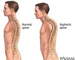
Abnormal curvatures in the human vertebral column must be observed in anatomical position. Excessive thoracic kyphosis is commonly known as hunchback or humpback. It is more commonly called Dowager's hump in older women as a result of osteoporosis, but it is also found in older men as well. The vertebrae erode and collapse causing a hump in the thoracic region and a loss in height. Excessive throracic kyphosis is characterized by an excessive posterior curve in the thoracic region.
Intervertebral disks are the cushions between the vertebral bodies. They are classified as symphyses joints and allow movement between individual vertebrae. IV discs act as shock absorbers for the vertebral column. IV discs contain an anulus fibrosus and a nucleus pulposus. The anulus fibrosus runs the outside of the IV disc and consists of fibrocartilage. The nucleus pulposus is considered the core of the IV disc. It contains mostly water and is held mostly accountable for the flexible nature of the IV disc. It is avascular, meaning, that it receives nutrients by diffusion from the anulus fibrosus and the verterbral body.
When there is a protrusion of the nucleus pulposus, it is often recognized as a hernia in the lower back. Herniation of the nucleus pulposus causes pain in the lower back as well as in the lower limbs. Although there are many causes of lower back pain, herniation of the nucleus pulposus is usually an asymptomatic finding. Most nucleus pulposus herniations occur in the lumbar and lumbosacracal region. This is due to the IV discs being the largest in this region and have the greatest amount of movement.